

Interactive CT & MRI Anat.Lite

وصف لـInteractive CT & MRI Anat.Lite
★Lite version★
This is the free Lite version of "Interactive CT and MRI Anatomy".
The function is restricted.
You can only see the transverse CT images of the head.
Please check the operation before purchasing the full version.
★ Details ★
This application is developed for medical students, interns, residents, doctors, nurses, and radiology technicians to understand the essential anatomical terms of the body.
You can learn anatomy by answering the terms by step-to-step questions using a total of 241 CT and MRI images.
A total of 17 images of 3D-CT, MRA and plain X-ray film(particularly the extremities) are included as references.
Other reference images include coronary artery segments defined by the American Heart Association(AHA), pulmonary segments, and liver segments(according to Couinaud classification).
You can enlarge all the images by simple manipulation.
★ Major functions ★
There are 4 major functions.
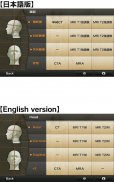
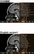
-1) Anatomical mode
Anatomical terms are overlaid on the images.
It can be used as the anatomical atlas.
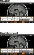
-2) Quiz mode type 1
You select the part of the image by using anatomical term.
Questions will basically appear randomly.
-3) Quiz mode type 2
You select the anatomical term by the part of the image.
Questions will basically appear randomly.
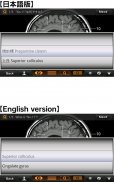
-4) Index
You can find the specific images by using anatomical terms.
★ Intended users ★
-Medical students
-Interns and residents
-Doctrors
-Nurses
-Radiology technicians
-All those who are intrested in CT and MRI anatomy
★ Images(a total of 258 images) ★
Images basically include horizontal, coronal, and sagital planes.
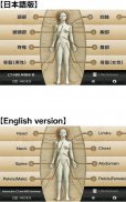
-Head(36 images including CTA and 3D-CT)
-Neck(24 images)
-Spine(19 images including plain X-ray films)
-Chest(61 images including 3D-CT images)
-Abdomen (37 images)
-Pelves: male (9 images)
-Pelvis: female (11 images)
-Extremities (shoulder, hand, elbow, hip joint, knee, foot) (61 images including plain X-ray films)
Editors
Toshiaki Nitori, M.D. (Professor of Radiology, Kyorin University, School of Medicine)
Yasuo Sasaki, M.D. (Manager of diagnostic radiology, Iwate Prefectural Central Hospital)
</div> <div jsname="WJz9Hc" style="display:none">★ ★ النسخة لايت
هذه هي النسخة لايت مجانا من "التفاعلية CT والرنين المغناطيسي التشريح".
يقتصر وظيفة.
يمكنك أن ترى إلا صورا CT عرضية من الرأس.
يرجى التحقق من العملية قبل شراء النسخة الكاملة.
★ ★ تفاصيل
تم تطوير هذا التطبيق لأسباب طبية الطلاب والمتدربين والمقيمين والأطباء والممرضين والفنيين الأشعة لفهم المصطلحات التشريحية الأساسية للجسم.
يمكنك معرفة التشريح عن طريق الإجابة شروط خطوة إلى خطوة الأسئلة باستخدام ما مجموعه 241 CT والرنين المغناطيسي الصور.
وشملت ما مجموعه 17 صور 3D-CT، MRA وفيلم عادي الأشعة السينية (وخاصة في الأطراف) كمراجع.
وتشمل الصور مرجعية أخرى قطاعات الشريان الذي حددته الرابطة الأمريكية لأمراض القلب (AHA)، وقطاعات الرئوية، وشرائح الكبد (وفقا لتصنيف Couinaud).
يمكنك تكبير جميع الصور عن طريق التلاعب بسيطة.
★ ★ الوظائف الرئيسية
هناك 4 وظائف رئيسية.
-1) وضع التشريحي
ومضافين حيث التشريحية على الصور.
ويمكن استخدامه كما الأطلس التشريحية.
-2) مسابقة نوع واسطة 1
تحديد جزء من الصورة باستخدام مصطلح التشريحية.
سوف تظهر الأسئلة أساسا عشوائيا.
-3) مسابقة نوع وضع 2
تحديد المدى التشريحي من قبل جزء من الصورة.
سوف تظهر الأسئلة أساسا عشوائيا.
-4) مؤشر
يمكنك العثور على صور محددة باستخدام المصطلحات التشريحية.
★ ★ المستخدمين المستهدفين
الطلاب -Medical
-Interns والمقيمين
-Doctrors
-Nurses
الفنيين -Radiology
* ضمان مشاركة جميع أولئك الذين مهتم في CT والرنين المغناطيسي التشريح
★ صور (أي ما مجموعه 258 صور) ★
صور تشمل أساسا الطائرات الأفقية، الاكليلية، وsagital.
-Head (36 الصور بما في ذلك CTA و3D-CT)
-Neck (24 صور)
-Spine (19 صورة بما في سهل أفلام الأشعة X)
-Chest (61 الصور بما في ذلك الصور 3D-CT)
-Abdomen (37 صور)
-Pelves: ذكر (9 صور)
-Pelvis: الإناث (11 صور)
-Extremities (الكتف واليد والكوع ومفصل الورك والركبة والقدم) (61 الصور بما في ذلك سهل أفلام الأشعة X)
المحررين
توشياكي Nitori، MD (أستاذ الأشعة بجامعة Kyorin، كلية الطب)
ياسو ساساكي، MD (مدير الأشعة التشخيصية، مستشفى ايواتي المحافظات الوسطى)</div> <div class="show-more-end">


